New Science: Studying Whale Sharks in the Galapagos with Underwater Ultrasound and Blood Sampling
We’re very happy to share a new scientific paper, published today in Endangered Species Research (free to read), that describes two shiny new research techniques: the use of underwater ultrasound to study the reproduction of free-swimming whale sharks, and how we can collect blood samples from these same whale sharks in the wild.

Dr Rui Matsumoto taking an ultrasound of an adult female whale shark at Darwin Island in the Galapagos
Honestly, it’s some mind-blowing stuff.
The whale shark is a relatively well-studied species… but ‘relative’ is key here. Only a single pregnant female, caught in a Taiwanese fishery back in 1995, has ever been examined by scientists – and we don’t know where the babies live, either. It’s mostly juvenile male sharks that frequent coastal areas, including tourism sites. Female whale sharks, both juveniles and adults, probably live further offshore; where they’re difficult to find, let alone study.
Darwin, the northernmost island in the Galapagos archipelago, is one of the only places we can consistently find large female whale sharks. Between June and October each year, there’s a good chance to see them swimming by the main dive site at the island, Darwin Arch, presenting a ‘huge’ opportunity to learn more about the whale shark life cycle.
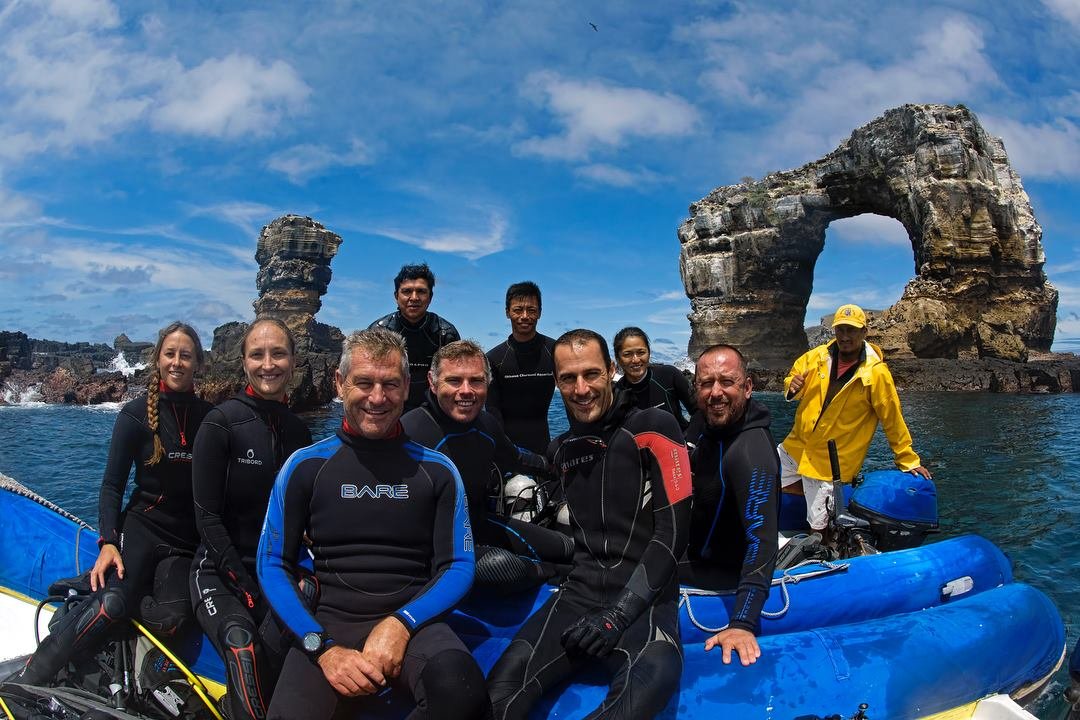
Some of the project team at Darwin Arch
Dr Rui Matsumoto, the lead author on this new paper, has been working with his colleagues at the Okinawa Churashima Foundation in Japan to build an underwater ultrasound and blood sampling kit. The Galapagos Whale Shark Project team have huge expertise at Darwin so, together, they developed an international project to test and refine these new methods on adult whale sharks in the wild. The team also included the Dirección Parque Nacional Galápagos, Universidad San Francisco de Quito, Kumamoto University, Massey University, Georgia Aquarium, along with Dr Chris Rohner and myself from MMF.
Applying these techniques was, unsurprisingly, not without some challenges. We were working at 10–30 m beneath the surface, often in strong current. Whale sharks are wonderfully placid, but they’re a lot faster than us, and emerge from the blue with little warning. We only had a short time to work with each animal. The ultrasound unit is the size of a large briefcase, so it’s far from hydrodynamic. Rui was using a ‘jetpack’ (an underwater propeller mounted on his air tank) to keep up with the sharks and scan them with the ultrasound wand, while filming the process with a GoPro to synchronize the ultrasound imagery with its position on the shark. (I felt cool by general association.)

Rui, with ‘jetpack’ 😎
Interpretation of the scans requires a lot of experience, too. Kiyomi Murakumo has been examining underwater ultrasound imagery in captive sharks and rays in the Okinawa Churaumi Aquarium for years now, monitoring pregnancy across species from nurse sharks to manta rays. She was able to discern the developing follicles within these greyscale images that prove that these female whale sharks are indeed adults – something that was previously only possible to determine through dissections.
Collecting blood samples from free-swimming sharks is, similarly, something that has taken a lot of trial and error to get right. Whale sharks have extremely thick skin, so we can’t reach their actual veins – instead, we’re trying to draw blood from vascularised tissue on their fins, which requires a large dose of luck. We also have to avoid contamination from seawater, which has necessitated the development of a two-syringe system, with one creating an initial vacuum, enabling the second to draw pure blood.
Dr Alistair Dove collecting a blood sample
When it was successful, though, it was possible to measure the levels of estradiol, progesterone and testosterone in wild, adult female whale sharks for the first time. In conjunction with the ultrasound, that allows us to finally start studying the whale sharks’ breeding cycle – all while the sharks swam blithely past, happily indifferent to our excitement.
This publication is just the start of that work, formally explaining the methods and their application to shark reproductive studies. There’s a ton more to do (including actually locating some of these aforementioned pregnant females). As we progress, it’ll allow for other research too, such as looking at stress and pollution levels in whale shark populations, thereby improving our understanding of their conservation needs.
Ultimately, we want to get to the point where we can do regular health check-ups on these endangered giants, identify the habitats that are most important to their life cycle, and ensure they’re well-protected. And if we have to use underwater jetpacks to accomplish that… well, I’d never call myself a hero, but people might say it about me. 😇
The full paper is here.
Learn more about the project on the MMF Galapagos Whale Shark Project page
See more media coverage:
ScienceNews. ‘Jet packs’ and ultrasounds could reveal secrets of pregnant whale sharks
MONGABAY. Uterine implants and underwater ultrasounds aim to demystify shark births
FORBES. Galapagos Whale Sharks Undergo Ultrasounds For First Time
Scuba Diver Magazine. Galapagos Whale Shark Research
DIVE Magazine. Whale shark reproductive ultrasound study published
–– Simon Pierce.
This study was made possible by a grant to the Galapagos Whale Shark Project from the Galapagos Conservation Trust, funding from the Planeterra Foundation to MMF, and the PADI Foundation. Chris and my contributions to this project were supported by Aqua-Firma, Shark Foundation, the Swiss Friends of Galapagos, Temperatio, and Waterlust. Thanks so much to all the GWSP and broader team for making this such a great project to work on!